PhD Symposium 2021 logo

The Big Picture
Zooming into Life

23rd EMBL PhD Symposium
16th - 17th December 2021
EMBL Venue | VIRTUAL


About the Symposium

Join us for the 23rd Anniversary of the annual EMBL PhD Symposium. Since its conception in 1999, the EMBL PhD Symposium has evolved into a highly respected scientific meeting, connecting young researchers and high-profile scientists alike. This year, our Symposium centers on the theme

“The Big Picture: Zooming into Life”

Each year, the life sciences are becoming more and more interdisciplinary in nature. No better example can be given than the current pandemic, where researchers in different fields have collaborated to bring about a rapid research-driven response against the novel coronavirus. We are dedicated to creating a symposium that brings together researchers who study life sciences at different scales and explore the interdisciplinary approaches utilized to link the different scales of life. We will address the most recent developments in the life sciences starting with taking a step back to look at life outside of Earth (astrobiology) and then proceed to zoom in covering ecology, organismal biology, tissue biology, cell biology and subcellular biology . Furthermore, we look to provoke fruitful discussion into how these topics can be integrated to promote better understanding of both the whole picture and its parts.
Speakers

A. Murat Eren (Meren) A. Murat Eren (Meren) Andi Pauli Andi Pauli Christoph Bock Christoph Bock Florian Schmidt Florian Schmidt Jan Brugues Jan Brugues Jennifer Gardy Jennifer Gardy Lynn Rothschild Lynn Rothschild Marie Manceau Marie Manceau Matthias Lutolf Matthias Lutolf Michael Levin Michael Levin Richard Wombacher Richard Wombacher Sara-Jane Dunn Sara-Jane Dunn Sarah Heilshorn Sarah Heilshorn Simon Haas Simon Haas Smita Shankar Smita Shankar Suliana Manley Suliana Manley Valentina Greco Valentina Greco
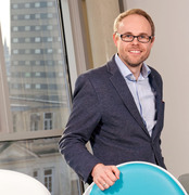
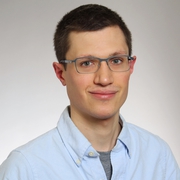
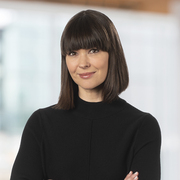
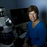
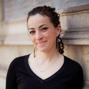
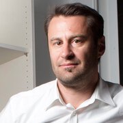
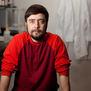
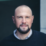
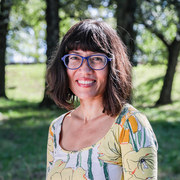
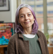
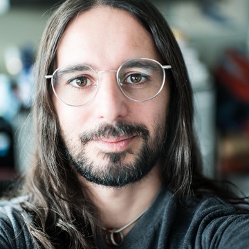
Photo of A. Murat Eren (Meren)

Dr. A. Murat Eren (Meren)

University of Chicago

A. Murat Eren (Meren) is a computer scientist and microbial ecologist who studies microbial lifestyles in a wide range of habitats that span from oceans and the human gut to sewage infrastructures and insect ovaries. Positioned at the unique intersection between computer science and microbiology, Meren and his group (https://merenlab.org) combine state-of-the-art computational strategies and molecular approaches to shed light on the ecology and evolution of naturally occurring microbial populations, with the primary aim of understanding strategies by which microbes thrive within their ever-changing environments. With their commitment to making science open and accessible, the members of the group are among the most active developers of anvi’o, an open-source software platform that strives to empower microbiologists by providing analytical tools and visualization strategies that aid in comprehending the avalanche of new data that is reshaping microbiology.
Photo of Andi Pauli

Dr. Andi Pauli

Research Insitute of Molecular Pathology
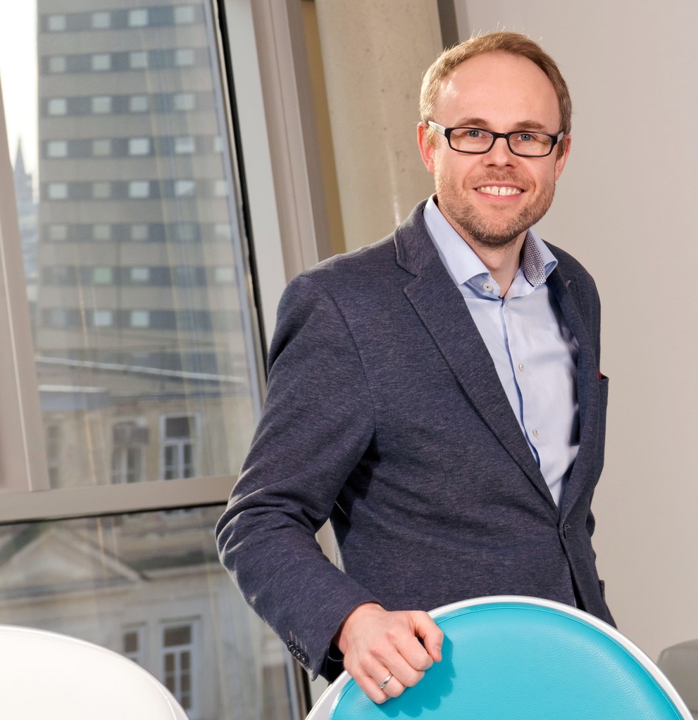
Andrea Pauli (Andi) studied biochemistry in Regensburg, Germany, and obtained her Masters in Molecular and Cellular Biology from Heidelberg University, Germany. In 2004, she started her PhD at the Research Institute of Molecular Pathology (IMP) in Vienna, Austria, co-supervised by Kim Nasmyth and Barry Dickson to investigate non-mitotic functions of cohesin using Drosophila as a model organism. In 2006, she moved with Kim Nasmyth to Oxford University, UK, where she obtained her PhD in 2009, providing the first direct evidence that cohesin has essential functions in post-mitotic cells. As a postdoc in Alex Schier's lab at Harvard University, USA, Andi made two key findings that have shaped her research since: first, translation is widespread outside of protein-coding regions in vertebrates; and second, some of the newly discovered translated regions encode functionally important short proteins, one of which is Toddler, an essential signal for mesodermal cell migration during gastrulation. In 2015, Andi established her own lab at the IMP in Vienna, Austria, with a current focus on (1) the mechanism of vertebrate fertilization and (2) translational regulation during the egg-to-embryo transition. The long-term vision of the Pauli lab is to unravel new concepts and molecular mechanisms governing this fascinating developmental transition that marks the beginning of life. Andi's work has been funded by EMBO, HFSP, the NIH grant to independence (K99), the FWF START Prize, and a Whitman Center Fellowship from the Marine Biological Labs. In 2018, Andi became an EMBO Young Investigator (EMBO YIP), and in 2021 she was elected as an EMBO Member.
Photo of Christoph Bock

Prof. Christoph Bock

CeMM Research Center for Molecular Medicine
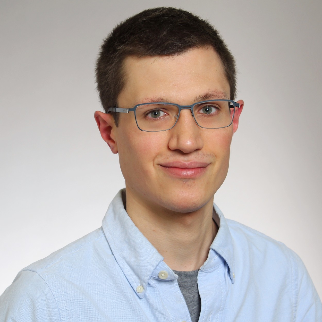
Christoph Bock is a Principal Investigator at the CeMM Research Center for Molecular Medicine of the Austrian Academy of Sciences and Professor of [Bio]Medical Informatics at the Medical University of Vienna. His research combines experimental biology (high-throughput sequencing, epigenetics, CRISPR screening, synthetic biology) with computational methods (bioinformatics, machine learning, artificial intelligence) – for cancer, immunology, and precision medicine. Before coming to Vienna, he was a postdoc at the Broad Institute of MIT and Harvard (2008-2011) and a PhD student at the Max Planck Institute for Informatics (2004-2008). Christoph Bock is also scientific coordinator of the Biomedical Sequencing Facility of CeMM and MedUni Vienna, informatics group leader at the Ludwig Boltzmann Institute for Rare and Undiagnosed Diseases (LBI-RUD), coordinator of an EU Horizon 2020 project that contributes single-cell sequencing of human organoids to the Human Cell Atlas, fellow of the European Lab for Learning and Intelligent Systems (ELLIS), and elected member of the Young Academy of the Austrian Academy of Sciences. He has received important research awards, including the Otto Hahn Medal of the Max Planck Society (2009), an ERC Starting Grant (2016-2021), an ERC Consolidator Grant (2021-2026), and the Overton Prize of the International Society for Computational Biology (2017).
Photo of Florian Schmidt

Dr. Florian Schmidt

ETH Zurich

Florian Schmidt is a postdoctoral fellow in the Platt group at the Department of Biosystems Science and Engineering (D-BSSE) at ETH Zurich. Florian obtained his B.Sc. and M.Sc. at the Ruprecht Karl University of Heidelberg where he entered the field of biology winning the 2013 iGEM competition together with fellow students as well as working on CRISPR/AAV gene therapy in the group of Dirk Grimm. During a short stint in the laboratory of Feng Zhang at the Broad Institute of MIT and Harvard, he helped to develop the first AAV-based CRISPR screening library delivered to a living mouse. Together with Randall Platt he then transitioned from Boston to Prof. Platt’s newly founded Laboratory for Biological Engineering at ETH Zurich where he pursued his predoctoral studies and established living microbial diagnostics based on the Record-seq technology he will present during this talk.
Photo of Jan Brugues

Dr. Jan Brugues

Max Planck Institute of Molecular Cell Biology and Genetics
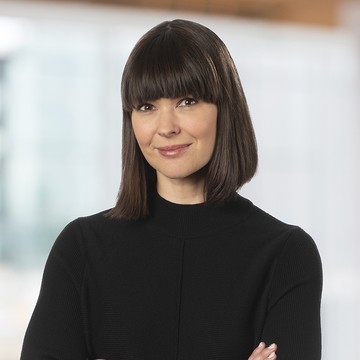
Jan Brugués is a jointly appointed Research Group leader at the MPI of Molecular Cell Biology and Genetics and the MPI for the Physics of Complex systems in Dresden since 2013. His lab works at the interphase between soft matter physics, cell biology, and theory to uncover the principles of how cellular compartments emerge from the interaction of individual molecules. Before starting his research group, he was trained as a theoretical physicist during his PhD jointly between ESPCI Paris (France) and University of Barcelona (Spain). As an HFSP at Harvard University, he investigated how mitotic spindles self-organize from local microtubule interactions despite their high turnover. In his lab, they complement quantitative measurements in vivo with reconstitution approaches to rebuild cellular functions using cell extracts and purified components. They combine these approaches with the development of new quantitative biophysical methods and theory. The goal of the Brugués lab is not only to understand particular cellular processes, but to identify general principles of protein self-organization to eventually provide the physical basis of how cells work. To achieve this, research in his group is centered around two general questions, both aiming at understanding the emergence of cellular compartmentalization. First, they want to understand how the size and shape of spindles arise from the interplay of mechanics, microtubule nucleation, and motor activities, and how these properties change during early development. Second, they want to understand the principles that govern chromatin organization in the nucleus, a question that has been largely ignored from the physical point of view. Both research directions synergize and benefit from their strong expertise in theory, quantitative microscopy, biophysical approaches, and reconstitution.
Photo of Jennifer Gardy

Dr. Jennifer Gardy

Bill & Melinda Gates Foundation

Dr. Jennifer Gardy joined the Bill & Melinda Gates Foundation’s Malaria team as Deputy Director, Surveillance, Data, and Epidemiology in February 2019. Before that, she spent ten years at the BC Centre for Disease Control and the University of British Columbia’s School of Population and Public Health, where she held the Canada Research Chair in Public Health Genomics. Her research focused on the use of genomics as a tool to understand pathogen transmission, and incorporated techniques drawn from genomics, bioinformatics, modelling, information visualization, and the social sciences. In 2018, Jennifer was named one of BC’s Most Influential Women in STEM by BC Business Magazine and was named one of the Government of Canada’s 20 Women of Impact in STEM. In addition to her science work, Jennifer is also an award-winning science communicator, hosting many episodes of science documentary television, including The Nature of Things and Daily Planet, as well as authoring science books for children, including a new book to be released in 2021.
Photo of Lynn Rothschild

Dr. Lynn Rothschild

NASA Ames Research Center

Lynn Rothschild, a research scientist at NASA Ames and Adjunct Professor at Brown University, is passionate about the origin and evolution of life on Earth and elsewhere, while at the same time pioneering the use of synthetic biology to enable space exploration. Her research focuses on how life, particularly microbes, has evolved in the context of the physical environment, both here and potentially elsewhere. She has brought her imagination and creativity to the burgeoning field of synthetic biology, articulating a vision for the future of synthetic biology as an enabling technology for NASA’s missions, including human space exploration and astrobiology. Since 2011 she has served as the faculty advisor of the Stanford-Brown iGEM team. Her lab is testing these plans in space on in the PowerCell secondary payload on the DLR EuCROPIS satellite. A past-president of the Society of Protozoologists, she is a fellow of the Linnean Society of London, The California Academy of Sciences and the Explorer’s Club. She was awarded the Isaac Asimov Award from the American Humanist Association, and the Horace Mann Award from Brown University. She has been a NASA Innovative Advanced Concepts (NIAC) fellow four times. Lynn was formerly Professor (Adjunct) at Stanford where she taught “Astrobiology and Space Exploration” for a decade.
Photo of Marie Manceau

Dr. Marie Manceau

College de France

Marie is interested in studying the formation and evolution of patterns in the skin. She completed her PhD in avian developmental biology in the laboratory of Pr. Marcelle at the University of Marseille (France) in 2007, and then moved as a postdoc in the laboratory of Dr. Hoekstra at Harvard University, where she studied the developmental bases of color pattern variation in rodents. Since 2013, she is a Young Research Group Leader at the College de France (CIRB). She also works a few weeks a year as a naturalist guide in the Arctic and Antarctica.
Photo of Matthias Lutolf

Prof. Matthias Lutolf

École polytechnique fédérale de Lausanne (EPFL)
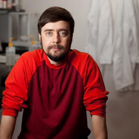
Professor Matthias Lutolf is Director of the Laboratory of Stem Cell Bioengineering at École polytechnique fédérale de Lausanne (EPFL), Switzerland. His highly innovative and cross-disciplinary research program is focused on the development of bio- and tissue-engineering strategies for improving organoid culture and enabling its translation to real-life applications.
Photo of Michael Levin

Dr. Michael Levin

Tufts University

Michael Levin’s original background was in computer science and software engineering, and he worked in artificial intelligence and scientific computing. He received his PhD in molecular genetics from Harvard, uncovering the genetic pathway underlying invariant left-right asymmetry of vertebrate embryogenesis. He did post-doctoral work at Harvard Medical School identifying long-range physiological communication upstream of the laterality transcriptional cascade. He started his independent lab at Forsyth Institute in 2000, developing the first molecular tools for probing endogenous bioelectrical signaling in embryogenesis. In 2009, he moved his lab to Tufts University, and in 2016 became the director of the new Allen Discovery Center at Tufts University. His group works at the interface between developmental biology, cognitive science, and computer science. Using biophysics, computational modeling, and molecular genetics, the Levin lab investigates the information processing in tissues that enables cells to cooperate towards the self-assembly and repair of complex anatomies. Developing mathematical models and novel ion channel modulation strategies, they work to understand how all cell types, not just neurons, form electrical networks that make decisions about growth and form. Applications have included reprogramming organ identity, inducing regeneration of appendages, normalizing tumors, and repairing of brain and craniofacial birth defects. By re-writing the bioelectrical “pattern memories” in tissues, they seek to develop applications for regenerative medicine, birth defects, cancer, and synthetic morphology.
Photo of Richard Wombacher

Dr. Richard Wombacher

Institute of Pharmacy and Molecular Biotechnology, Heidelberg
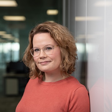
Richard Wombacher is group leader at the Chemical Biology Department of the Max-Planck-Institute for Medical Research in Heidelberg. Richard graduated in chemistry at the Free University of Berlin and received his Ph.D. at the Ruprecht-Karls University working in the field of RNA catalysis. After a post-doc with Virginia Cornish at the Columbia University in New York, he started his own research group at the Ruprecht-Karls University, working in in the field of chemical biology and protein chemistry. In 2020 Richard became group leader at the MPI for Medical Research in Heidelberg. The research of his group is focused on the development and application of chemical tools for control of protein function and modification. This includes biorthogonal chemistry, probe development for advanced imaging technologies like super-resolution microscopy and single molecule microscopy as well as optochemicals for protein manipulation with high spatiotemporal control.
Photo of Sara-Jane Dunn

Dr. Sara-Jane Dunn

Google DeepMind

Dr Dunn is a Research Scientist at DeepMind. She has a Masters in Mathematics from the University of Oxford, where she also achieved her doctorate in Computational Biology. Prior to DeepMind she was a Principal Scientist at Microsoft Research, having joined as a Postdoctoral Researcher in 2012. She is an Affiliate PI of the Wellcome-MRC Stem Cell Institute, University of Cambridge. Her research interests are in the domain of biological computation: how we can uncover the fundamental principles of biological information-processing, and use this understanding to tackle important, impactful problems in the field.
Photo of Sarah Heilshorn

Prof. Sarah Heilshorn

Stanford University

Heilshorn's interests include biomaterials in regenerative medicine, engineered proteins with novel assembly properties, microfluidics and photolithography of proteins, and synthesis of materials to influence stem cell differentiation. Current projects include tissue engineering for spinal cord and blood vessel regeneration, designing injectable materials for use in stem cell therapies, and the design of microfluidic devices to study the directed migration of cells (i.e., chemotaxis).
Photo of Simon Haas

Dr. Simon Haas

Heidelberg Institute For Stem Cell Technology And Experimental Medicine
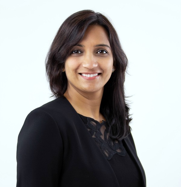
As of 2020, Simon Haas is an independent group leader at the Berlin Institute of Health (BIH), the Charité university medicine and the Berlin Institute of Medical Systems Biology (BIMSB) at the Max Delbrück Center (MDC) in Berlin, Germany. He is also an associated group leader at the German Cancer Research Center (DKFZ) and the Heidelberg Institute for Stem Cell Technology and Experimental Medicine (HI-STEM gGmbH), Heidelberg, Germany. Simon studied Molecular Cell Biology, Biochemistry and Molecular Biosciences at Heidelberg University, Imperial College London and the DKFZ. He received his PhD from DKFZ and Heidelberg University in 2016 (with Marieke Essers). Simon performed research at DKFZ, Massachusetts Institute of Technology (MIT) and Harvard. Since 2016, he is a junior group leader at HI-STEM (with Andreas Trumpp). His research focus is to understand the blood and immune system, and how it contributes to disease, in particular cancer. For this purpose, his lab develops single-cell transcriptomic, genomic, surface proteomic, functional and spatial technologies, and combines those with classical approaches from the fields of immunology, stem cell biology and cancer. As part of the hematology, oncology and tumor immunology departments of Europe’s largest university hospital at the Charité and core member of the BIH / MDC focus area ‘Single-Cell Approaches for Personalized Medicine’ one of his main goals is to transform single-cell technologies into precision oncology approaches that provide ultra-precise diagnostics, prognostics and guide therapy choice in cancer therapy.
Photo of Smita Shankar

Dr. Smita Shankar

Impossible Foods

Smita Shankar is the VP of Biomanufacturing at Impossible Foods. She leads the team responsible for manufacturing and scaling Impossible Burger’s “magic” ingredient – soy leghemoglobin, or “heme” – which provides the unmistakable flavor and aroma of meat. Previously, Smita was the VP of R&D at Impossible. Smita joined Impossible Foods in 2013 as one of the company’s first R&D hires. Following Impossible’s discovery of heme as a key ingredient in 2014, she built the company’s microbial strain development program from scratch, leading to industrial-scale production of recombinant proteins. Over the next seven years, Smita oversaw the implementation of new research and technology, built multiple production plants, and established a world class team of scientists and engineers. In 2017, her work resulted in a mission-critical patent covering Impossible Foods’ unique method of producing heme at scale. Prior to Impossible Foods, Smita worked at Codexis engineering microbes to make sustainable bio-based detergent alcohols.Smita got her Ph.D. in Biochemistry, Molecular and Cellular Biology at Cornell University, where she studied bacterial gene expression. Following her Ph.D. she was a postdoctoral fellow at the University of California, San Francisco, where she researched the genetic manipulation of yeast through a fellowship from the Leukemia and Lymphoma Society.
Photo of Suliana Manley

Dr. Suliana Manley

Ecole polytechnique federale de Lausanne
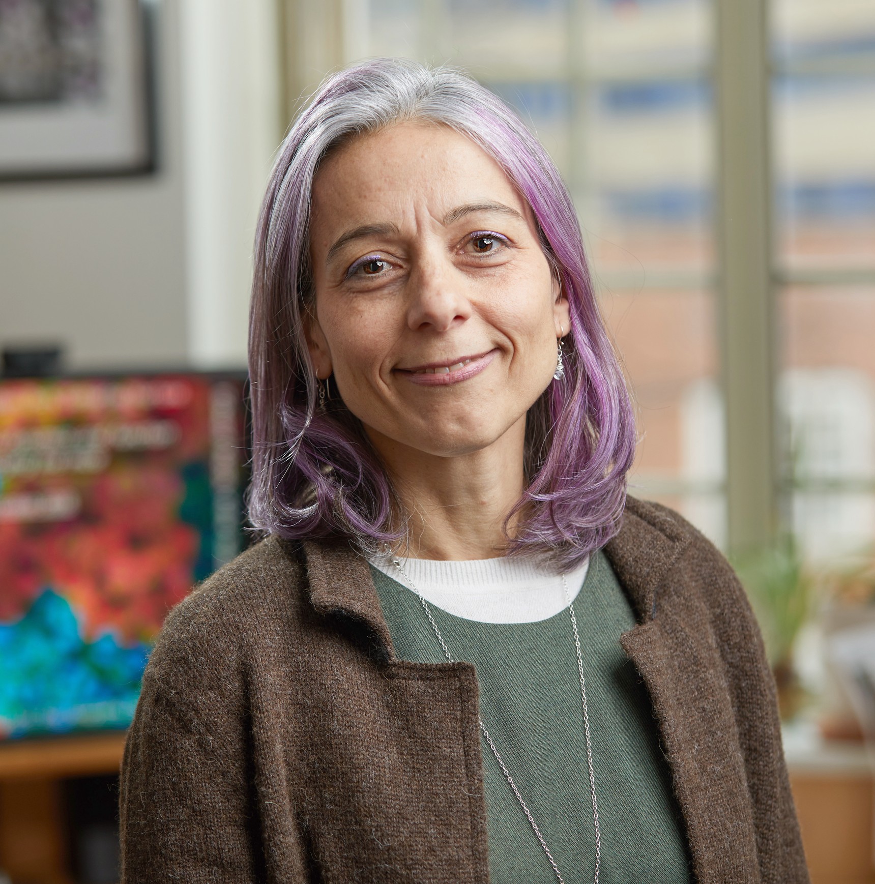
Born in the United States, Suliana Manley studied at Rice University where she obtained a bachelor's degree in physics and mathematics in 1997, cum laude. She continued her studies at Harvard University, where she earned a PhD in physics under the supervision of Prof. Dave Weitz in 2004. After this, she conducted postdoctoral research on lipid bilayer and red blood cell membrane dynamics at MIT. She subsequently went on to work as a postdoctoral researcher in the cell biology laboratory of Jennifer Lippincott-Schwartz at the National Institutes of Health (USA). During this time, she developed a highly promising optical method (sptPALM) for studying the dynamics of large ensembles of single proteins in membranes and inside cells. Currently, at the Ecole polytechnique federale de Lausanne, her lab develops and utilizes automated super-resolution fluorescence imaging techniques combined with live cell imaging and single molecule tracking to determine how the dynamics of protein assembly are coordinated.
Photo of Valentina Greco

Dr. Valentina Greco

Yale School of Medicine

Valentina Greco, PhD, is the Carolyn Walch Slayman Professor of Genetics at Yale University. She studied Molecular Biology at the University of Palermo (Italy), and later pursued her PhD at EMBL/MPI-CBG. After a postdoc at the Rockefeller University (USA), she joined the Yale Stem Cell Center, the Yale Cancer Center, and the Genetics, Cell Biology, and Dermatology Departments at Yale University, where she opened her lab. The Greco lab aims to define how tissues maintain themselves throughout our lives in the face of continuous cellular turnover, frequent injuries and spontaneous mutations. The lab also operates at the forefront of technological innovation, having established multiple approaches to visualize and manipulate stem cells in intact animals. During the course of her career, Dr. Greco has been honoured with several awards, including most recently the 2021 International Society for Stem Cell Research (ISSCR) Momentum Award, the 2019 NIH Director’s Pioneer Award, the 2019 Yale Postdoctoral Mentoring Award, the 2018 Yale Graduate Mentor Award in the Natural Sciences, and in 2018 she was named the Inaugural Holder of the Carolyn Slayman Endowed Professorship.

Programme

Wed
Thu
Fri

Wednesday, December 15th

13:00 - 14:00 Nikon
From millimeters to nanometres; Life seen through Nikon microscopes
14:00 - 15:00 Bayer
Part I: Why is science so important for society? Equality, Collaboration & Trust.
Part II: Careers in Science & Beyond: A Real World Experience
15:00 - 16:00 Agilent
From Academia to Industry: Transforming the labs that transform the world.
15:00 - 17:00 Virtual Booths
Bayer and Agilent

Thursday, December 16th

08:45 - 09:00 Symposium Opening & Welcome: Edith Heard

Session 1 - Subcellular

9:00 - 9:05 Subcellular Session Introduction
9:05 - 9:35 Richard Wombacher: Chemical probes for application in biology and medicine
9:35-10:05 Suliana Manley: Mysteries and insights into mitochondrial dynamics
10:05-10:35 Jan Brugues: Force generation by protein-DNA co-condensation
10:35-10:55 Short Talks
Carmen Escalona - Non-viral CRISPR delivery strategies for the generation of improved gene editing tools
Sudarshana Laha - Phase separated compartments as biochemical reactors
Michael Jendrusch - AlphaDesign - designing proteins with AlphaFold
10:55-11:10 Sponsor Talks: Bayer, Nikon, and Agilent

Poster Session

11:10-11:15 Virtual Coffee Break
11:15-12:00 Posters A-J
12:00-13:00 Lunch

Session 2 - Cell

13:00-13:05 Cell Session Introduction
13:05-13:35 Florian Schmidt: Transcriptional recording by CRISPR spacer acquisition from RNA
13:35-14:05 Simon Haas: Stem cells & cancer at single-cell resolution
14:05-14:25 Short Talks
Elzbieta Gralinska - What do cells have in common? Association Plots reveal cell-cluster specific genes from high-dimensional transcriptome data
Sahana Vasudevan - Eavesdropping attack of bacterial communication by functionalized metal oxide nanointerfaces: Towards early infection diagnosis
Craig Watson - Microfluidic chip for long-term cell co-culture
14:25-14:35 Virtual Coffee Break
14:35-15:05 Christoph Bock: Looking into the past and future of cells: Single-cell analysis of epigenetic cell states in cancer and immunology
15:05-15:35 Sara-Jane Dunn: From Software Bugs to Transcriptional Networks - Uncovering Information Processing at the Cellular Level
15:35-16:05 Smita Shakar: Making Heme: Impossible Foods’ Key Ingredient

Workshops (see details)

16:05-16:20 Virtual Coffee Break
16:20-18:20 Workshops
Consultancy - Answering clients' problematics in innovation at the interface between science and business, Stephane bagnato
Career Panel - Sander Wuyts, Judith Zaugg, Maria Polychronidou, Mark Bonyhadi and Maciej Lopatka
Mental Health - How to become more productive, confident and happier researcher ?, Ewa Pluciennicka
Science Communication - Science journalists are not the enemy, Luca Barone
Bio-imaging center - Cryo-CLEM and Super-resolution methods, Simone Mattei and Timo Zimmermann
* Digital posters and pre-recorded short talks as well as discussion channels will be available for registered participants throughout the whole virtual meeting.
* All times in the programme below are shown as the time in Europe/Berlin. To find out the equivalent time zone in your location, enter Berlin, the programme time and date along with your city into the Time Zone Converter.

Friday, December 17th

Session 3 - Tissue

8:55-9:00 Tissue Session Introduction
9:00-9:30 Aidan Gilchrist (Sarah Heilshorn's lab): Engineered matrices probe essential features of environment across a broad spectrum of organoid systems
9:30-10:00 Marie Manceau: Developmental control of avian skin patterns
10:00-10:30 Matthias Lutolf: Engineering organoid development
10:30-10:45 Short Talks
Marina Cuenca - The role of cephalic furrow formation in Drosophila: From cellular forces to tissue flow
Alan Chramiec - Recapitulation of organ-specific breast cancer metastasis using an engineered multi-tissue platform

Poster Session

10:45-11:00 Virtual Coffee Break
11:00-11:45 Posters K-Z
11:45-12:30 Lunch
12:30-13:30 Social Activities

Session 4 - Organism

13:30-13:35 Organism Session Introduction
13:35-14:05 Michael Levin: Endogenous bioelectrical networks instruct growth and form: from pre-neural mechanisms to electroceuticals for regenerative medicine
14:05-14:35 Andi Pauli: A molecular network of conserved factors keeps ribosomes dormant in the egg
14:35-15:05 Valentina Greco: Principles of regeneration captured by imaging the skin of live mice
15:05-15:25 Short Talks
Salvo Danilo Lombardo - Common topological and biological network properties across different species
Mayra Martinez-Lopez - In vivo imaging of BCG immunotherapy reveals that macrophages play an essential role in tumor rejection
Fangwen Zhao - Chromatin profiling uncovers a gene-regulatory atlas underlying diverse monogenic antibody deficiencies
15:25-15:40 Virtual Coffee Break

Session 5 - Ecology

15:40-15:45 Ecology Session Introduction
15:45-16:15 Murat Eren: Studying the ecology of microbes through 'omics
16:15-16:45 Jennifer Gardy: Data-driven decision-making for malaria eradication
16:45-17:05 Short Talks
Lara Urban - Leveraging adaptive sampling of environmental DNA for monitoring the critically endangered kakapo
Hugo Barreto - Gut mélange à trois: fluctuating selection modulated by microbiota, host immune system, and antibiotics
Luke Steller - The origins of life on earth: a messy chemistry approach

Session 6 - Astrobiology

17:20-17:25 Astrobiology Session Introduction
17:25-17:55 Lynn Rothschild: Are we alone? The search for life in the Universe

Panel Discussion

18:00-19:00 Panel Discussion: Jan Brugues, Christophe Bock, Sarah Heilshorn, Valentina Greco, Jennifer Gardy, Lynn Rothschild

Closure

19:00-19:30 Closing Remarks and Awards for Best Talk (sponsored by Biology Open) & Best Poster (sponsored by Nikon)
* Digital posters and pre-recorded short talks as well as discussion channels will be available for registered participants throughout the whole virtual meeting.
* All times in the programme below are shown as the time in Europe/Berlin. To find out the equivalent time zone in your location, enter Berlin, the programme time and date along with your city into the Time Zone Converter.

Workshops

Photo of Herman Fennema Nikon Photo of Karl Collins Bayer Foundation Photo of To be announced Agilent Photo of Sander Wuyts, Judith Zaugg, Maria Polychronidou, Mark Bonyhadi, Maciej Lopatka Career panel Photo of Stephane bagnato Consultancy Photo of Ewa Pluciennicka Mental health Photo of Luca Barone Scientific communication Photo of Simone Mattei & Timo Zimmermann Imaging Centre
Photo of Simone Mattei & Timo Zimmermann

Bio-Imaging
16th December, 4.20-6.20 pm

Simone Mattei & Timo Zimmermann, EMBL Imaging Centre, Germany
The EMBL Imaging Centre represents a new EMBL service unit for cutting-edge EM and LM technologies and for the correlative approaches that combine the two imaging modalities. The main mission of the EMBL Imaging Centre is to rapidly make the most advanced microscopy technologies available to the international user community from both academia and industry to enable new ground-breaking research that crosses the scales of biology.
We provide users with access to a powerful and synergistic portfolio including academically developed methods not yet commercially available, we support their projects from sample preparation to image analysis, and we offer tailored training opportunities to users and imaging service facility managers.

The Imaging centre workshop is divided into 2 parts:

WORKSHOP A: "Cryo-CLEM"
Light microscopy allows the observation and analysis of biological processes inside live and fixed biological samples. Electron microscopy on the other hand can deliver structural information and the context in which they are embedded. Correlative light and electron microscopy (CLEM) combines information from both imaging modalities, allowing precise localization and targeting of rare events or heterogenous populations. Cryo-CLEM allows an in-depth understanding of cellular structural biology in their native environment, after preparing the sample in a close to native state. In this workshop you will learn how you can combine cryo-confocal imaging, cryo-lamella preparation and cryo-electron tomography to get high-resolution information of your favourite structure. The correlative workflow connects a confocal fluorescence microscope, with a focused ion beam - scanning electron microscope for targeted lamella preparation and a high-end transmission electron microscope. You will learn about the hardware and software involved to investigate your samples in their native state. During the workshop you will be able to talk to Embl scientists running those workflows within their daily routine offering those workflows as a service. You can receive practical information, troubleshooting advice and feedback about accessibility of those techniques.

WORKSHOP B: "Super-resolution methods"
After having been limited by the diffraction limit as stated by Ernst Abbe for many decades, light microscopy has recently overcome this barrier and is now able to image structures at unprecedented levels of detail. In this workshop we aim to provide a practical introduction into several of these methods for super-resolution light microscopy. We will cover one coordinate-targeting approach (Stimulated Emission Depletion Microscopy, STED), one based on stochastically detected single molecules (Single Molecule Localization Microscopy, SMLM) and a combination of both concepts that currently achieves the highest possible resolution (Minimal Photon Fluxes, MINFLUX). These methods are now openly available at the EMBL Imaging Centre as a service for researchers who want to use them in their experiments.
The different super-resolution methods work at different levels of resolution and offer different advantages and disadvantages in sample preparation requirements, labelling densities, sources of error and in speed and throughput. The workshop will accordingly cover what kind of methods are suitable for which experimental question, sample preparation considerations and on-site demonstrations of the image acquisition methods.
Photo of Luca Barone

Science journalists are not the enemy
16th December, 4.20-6.20 pm

Luca Barone, EMBL Barcelona
Science and the scientific endeavour play an increasingly central role in our society. Public policies are (or should be) informed by scientific advice, citizens have the right and the duty to take informed decisions about the science that pervades every aspect of our lives. But do we guarantee that the right information and the contextualized science gets to the general public? What role do scientists, press offices, journalists play in the transmission belt that guarantees that the discoveries made in the labs reach a broader audience? How do we guarantee that the information is accurate and its implications are not distorted?

We will play a little bit with some press releases, analyze their spin and observe what happens to the titles of the articles in the press.
Photo of Ewa Pluciennicka

How to become a more productive, confident and happier researcher?
16th December, 4.20-6.20 pm

Ewa Pluciennicka, PhDsuccess
The Mental Health of early career researchers is alarming. Several recent studies reported that PhD candidates are at a high risk of developing mental health disorders such as depression, anxiety, and burn-out (Evans, 2018).
The most common reasons for that are the excessive workload, stress, high expectations of academia, and lack of life-work balance (Leveque, 2017)
Despite the growing research in the field of mental health of researchers, little attention has been given to proposing practical solutions to improve the well-being of PhD researchers (Satinsky et al. 2021).

Thus, the aim of this training is to:
1. Raise mental health awareness among young researchers so they can be more conscious of the risk for their mental health and effectively avoid them.
2. Propose a practical solution on how to better organise their time and attention, so they can achieve more in a shorter time and consequently, reduce their stress level daily.
3. Highlight the importance of self-satisfaction and its impact on different areas of life and general well-being.

To achieve that, besides the theoretical explanations, a series of practical tools and self-reflection exercises will be shared. Finally, all the participants will be invited to discussion and experience sharing
Photo of Stephane bagnato

Answering clients' problematics in innovation at the interface between science and business
16th December, 4.20-6.20 pm

Stephane bagnato
In this session, you will be in the shoes of a consultant in innovation. With the other participants in your subgroup, you’ll be confronted with a client’s problem, and use your problem-solving skills to decompose the problem and think of how you can answer the question. You will collaborate to think of clever ways to answer the question. The world is yours, you will have access to your own knowledge, online tools, google search, etc. You will combine your ideas, perform an analysis and draft a recommendation to solve the initial problem. In the end, the members will elect a speaker in the group and pitch their idea as if the other members were clients, to give your analysis results and your recommendation.
Photo of Sander Wuyts, Judith Zaugg, Maria Polychronidou, Mark Bonyhadi, Maciej Lopatka

Career panel
16th December, 4.20-6.20 pm

Sander Wuyts, Judith Zaugg, Maria Polychronidou, Mark Bonyhadi, Maciej Lopatka
Five successful men and women of various scientific areas will share their experience with you through short presentations but most importantly want to answer your questions about career path and more. With us will be:
- Sander Wuyts, who created his own start-up: Immunowatch,
- Judith Zaugg, a group leader at EMBL Heidelberg,
- Maria Polychronidou, a senior editor of Molecular Systems Biology,
- Mark Bonyhadi, who after a scientific career in Biotechnology industry moved into venture capitalism,
- Maciej Lopatka, who participates in shaping the future of our countries through policy making regarding research and innovation.
Photo of To be announced

From Academia to Industry: Transforming the labs that transform the world.
15th December, 3-4 pm
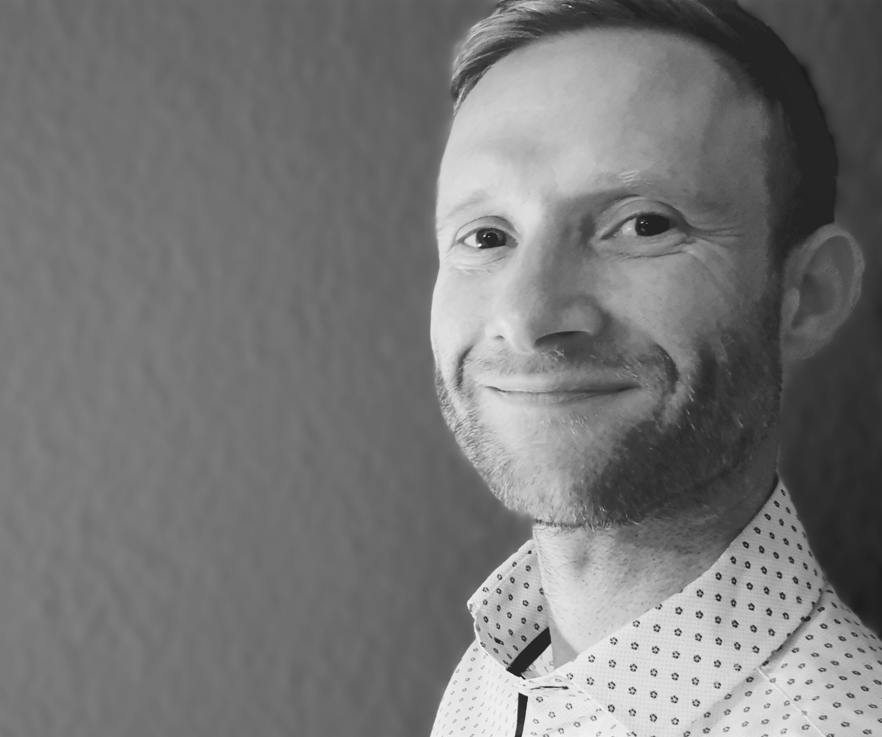
To be announced, Agilent
To be announced
Photo of Karl Collins

To be announced
15th December, 2-3 pm
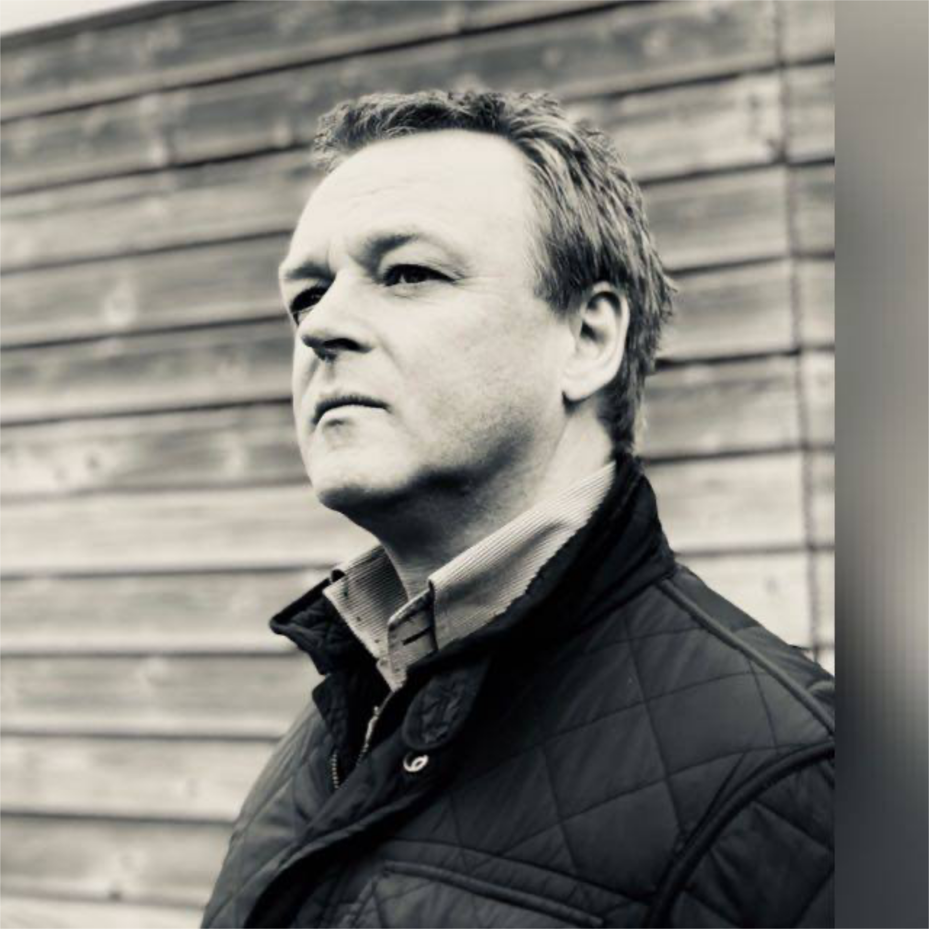
Karl Collins, Bayer Foundation
Originally from the north of England, Karl studied chemistry at the University of Manchester where he stayed to undertake his PhD in Synthetic Chemistry with Prof. David Procter. Karl then moved to Germany in 2012 for a research stay at the University of Münster in the Group of Prof. Frank Glorius, where he worked to develop novel metal catalysed chemical reactions and established methods to evaluate the real world application of synthetic chemistry. In 2014, he joined Bayer AG working as lab head in medicinal chemistry with a focus on cardiovascular diseases and where he also continued to develop his interest and expertise in high-through put experimentation & automation. Since January 2021, Karl has been responsible for the scientific portfolio of the Bayer Foundation with a strategic focus on driving equality, collaboration & trust in science.

In other news: Karl likes riding his bike, works actively to address gender inequality in science, has worked as a freelance scientific journalist, worked in schools across the UK to bring science to the next generation, sits on a variety of university advisory boards and related panels, and he likes organising conferences.

The workshop will cover two topics:
Part I: Why is science so important for society? Equality, Collaboration & Trust.
Part II: Careers in Science & Beyond: A Real World Experience
Photo of Herman Fennema

From millimeters to nanometres; Life seen through Nikon microscopes
15th December, 1-2 pm

Herman Fennema, Nikon
Life in all its forms can be seen all around us. Life in the microworld needs a microscope to visualize. In this presentation, we will take you through various sizes of life, ranging from watching the heartbeat of a Zebrafish in real time, all the way down to the movement of inner membranes of mitochondria, all the while illustrating what kind of microscopy hardware was used to obtain such images. We will also highlight how novel artificial intelligence applications help us produce an easier user experience and more powerful analyses and visualizations.

Registration

Registration is now closed.
Click to access late registration


Follow us on our social media accounts to stay tuned!

#EMBLTheBigPicture | #EMBLPhDSymp2021


FAQs

What is the EMBL PhD symposium about?

Answer: The EMBL PhD Symposium is organized by EMBL PhD students to serve as a gathering that connects young researchers and high-profile scientists alike. This year, our Symposium centers on the theme “The Big Picture: Zooming into Life”

When is the symposium taking place?

Answer: The symposium is scheduled to take place on the 16th and 17th of December 2021. In the evening before the symposium (15.12.21) we will offer an onboarding session to get in touch with each other and to get to know the platform.

Where is the symposium taking place?

Answer: The symposium will be virtual. The platform information will be shared closer to the symposium dates.

Who can attend the symposium?

Answer: Everyone is invited to attend the symposium.

How do I submit questions about the symposium?

Answer: Please feel free to contact phdsymposium2021@embl.de

How can I participate in the symposium?

Answer: You can participate in the symposium by registering at https://www.phdsymposium.embl.org

Can I submit an abstract for a talk and/or poster presentation?

Answer: Yes! All registrants are welcome to submit their abstracts. They can indicate if they want to give a talk or a poster presentation. The flash talks will be selected by the organizing committee after the submission deadline.

How do I submit an abstract?

Answer: Only registered applicants can submit abstracts. You can find the link for abstract submission in the confirmation email.

Do I need to submit an abstract to participate in the symposium?

Answer: While we encourage abstract submissions, we don’t require abstracts to participate in the symposium.

When do individuals with selected abstracts get to test the platform?

Answer: Participants with selected abstracts will be invited to test the platform before the symposium. The actual dates and times will be communicated accordingly.

When is the last day for registration for abstract and regular registration?

Answer: The last day for abstract submission is the 25th of October 2021. The last day to register as a regular participant is the 14th of November, 2021.

How much is the registration fee?

Answer: The registration fee is 25 euros for each registrant

What payment options are available?

Answer: There are two payment options available. You can either pay upon registration or choose the pay-later option.

Is there any financial assistance and how do I request financial assistance?

Answer: Yes we have financial assistance available for eligible participants. You can request assistance by visiting the abstract submission link and answering the questions marked with ‘Financial Assistance’. If you are not submitting an abstract, you can still apply for financial assistance by choosing ‘Financial Assistance’ and completing the aforementioned questions. The organizing committee will review the requests and select the recipients of registration fee waivers during the abstract selection process.

Sponsors

Sponsorship Information

The EMBL PhD symposium will provide an invaluable networking environment for your company to enhance the profile amongst the molecular biosciences research community, especially towards the young generation. The symposium will offer prime opportunities for your company to promote your new products, latest technology, and services. If you are interested in sponsoring our symposium and would like to have more information, direct your enquires to us.

Sponsors of the symposium

EMBL

Premium Sponsors

Nikon Agilent Bayer Foundation

Event Sponsors

Biology Open TRR83

Also kindly supported by:

EMBL CPP founder partners

Leica Microsystems Olympus

EMBL CPP corporate partners

10x Genomics BD Bio-Rad Boehringer Ingelheim Eppendorf Glaxo Smith Kline MACS-Miltenyi Biotec NetApp Stilla Technologies ThermoFisher Scientific

EMBL CPP associate partners

Merck New England Biolabs Nikon Promega Roche Sanofi Sartorius

Contact

Have a question? Please feel free to contact the PhD Symposium 2021 Organising Committee:
phdsymposium2021@embl.de

#emblphdsymposium

twitter facebook instagram